Edited by Grace Xiong, Harry Lightsey, Brendan Striano - 7/1/2021
Indications/Contraindications
Indications: Elbow dislocation
Contraindications: Ipsilateral injury (e.g. humerus or forearm fracture)
Technique (if your forearm is as long or longer than the patient’s forearm)
Materials: Splint - Have splint laid out in advance so that it can be applied once reduction is completed
Positioning:
Patient is upright or supine.
You may choose to have a second person hold countertraction, although with proper grip this can be done alone.
Technique:
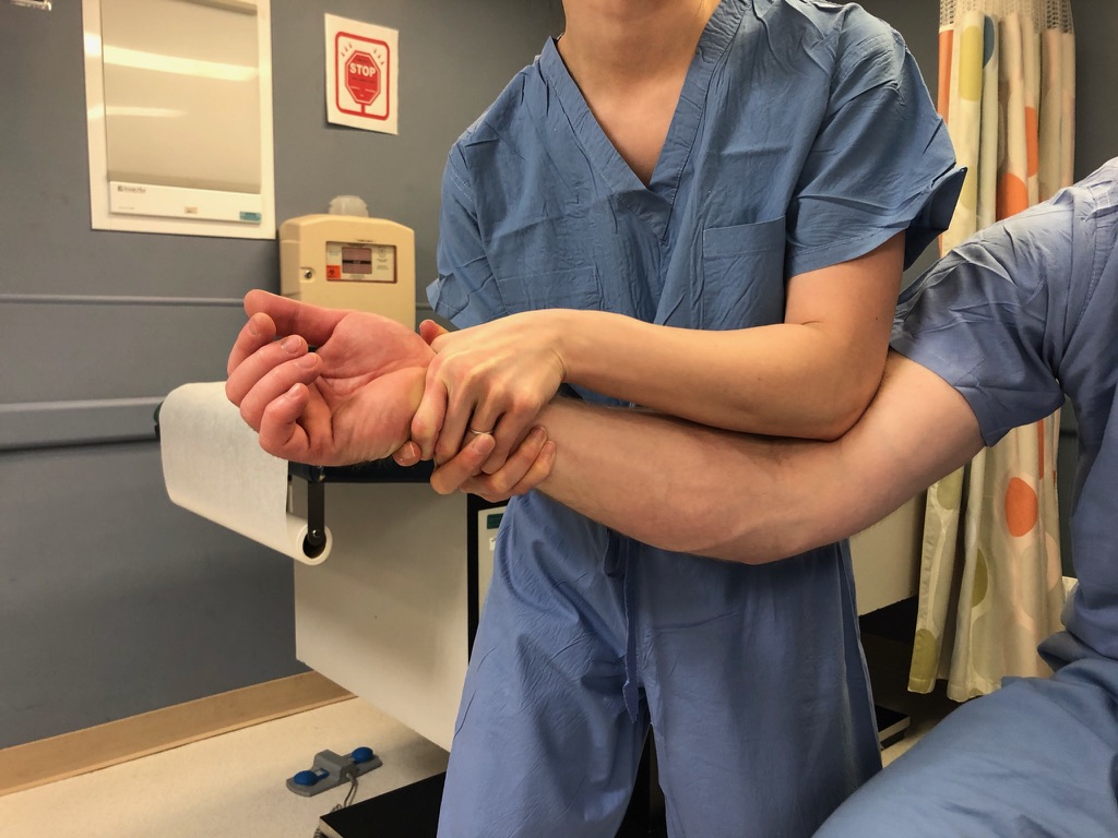
You face the patient and the arm that is farthest from the patient grips the antebrachial fossa/distal humerus to give yourself countertraction.
Your hand closest to the patient grips the hand/wrist of the patient.
Your hand closest to the patient then pulls in-line traction, then pulls the elbow into flexion while maintaining traction.
Once you feel a reduction, if you have fluoroscopy available, take the elbow through a range of motion (flexion/extension/pronation/supination).
Often the patient will be able to communicate to you whether the elbow is back reduced.
Splint with a posterior slab splint (very stable elbow) or posterior slab splint with side struts (moderately unstable) or double sugartong (very unstable).


Technique (if your forearm is shorter than the patient’s forearm)
Materials: None
Positioning:
Patient is upright or supine.
You may choose to have a second person hold countertraction, although with proper grip this can be done alone.
Technique: You stand on the side of the dislocation, with you facing the same direction as the patient (e.g., if the patient’s right elbow is dislocated, your left side is standing by their right side).
Grip the base of the patient’s wrist with the hand that is closest to the patient.
Place your olecranon into the antecubital fossa of the patient. Support this arm and stabilize with your other hand.
Pull in-line traction and flex at the same time, using your olecranon against the antecubital fossa as a fulcrum.
Once you feel a reduction, if you have fluoroscopy available, take the elbow through a range of motion (flexion/extension/pronation/supination).
Often the patient will be able to communicate to you whether the elbow is back reduced
Splint with a posterior slab splint (very stable elbow) or posterior slab splint with side struts (moderately unstable) or double sugartong (very unstable).

Post reduction imaging and protocols
Imaging: Post-reduction AP and lateral films of the elbow
Immobilization: Splint with a posterior slab splint (very stable elbow) or posterior slab splint with side struts (moderately unstable) or double sugartong (very unstable).
Restrictions: NWB upper extremity, encourage ROM at digits / shoulder (pendulums)
Follow up: 2 weeks ortho trauma, sports clinic, or upper extremity clinic
Pearls & Pitfalls
Potential complications:
Insufficient traction may cause the distal humerus to “scrape” over the coronoid as it reduces, causing iatrogenic injury.
Repeat dislocation can result if unstable elbows are allowed to range during the splinting process, or if the splinting is inadequate.
Tips for efficiency:
Have your set up before you begin (e.g. if you plan to splint, take fluoroscopy, etc., have all this turned on, at the bedside, rolled out, water ready, etc before you perform the reduction).
If you think the elbow is very unstable, enlist a second person to help you hold as you splint.