Edited by Grace Xiong, Harry Lightsey, Brendan Striano - 7/1/2021
Indications/Contraindications
Indications: Ankle Fracture Dislocation of any type (Reduction type will vary with the morphology of the fracture)
Contraindications: No contraindications to ankle reduction, but be thorough with XR evaluation so as to not perform the wrong reduction maneuver
Relative contraindications: None
Setup and Preparation
Consent: It is important to explain to the patient that this is an attempt at a reduction and that failure may mean a second reduction attempt. Additionally, explain the low, but potential risk of iatrogenic fracture or nerve injury
Technique
Materials:
Materials for Ankle Hematoma block
Splint Materials
Positioning:
Patient is supine on stretcher slid down towards the foot of the bed
Stretcher should be dropped as low as possible
Have the patient flex the hip and knee to 90 degrees and hold that position with their hands
Technique (Spiral Fibula Fracture with Transverse Medial Mal Fx or Intact Medial Mal w/ Wide Medial Clear Space):
Perform ankle block first
While block kicks in, roll out the splint
If the talus is posteriorly dislocated, place one hand posteriorly on the calcaneus and one hand anteriorly on the tibia
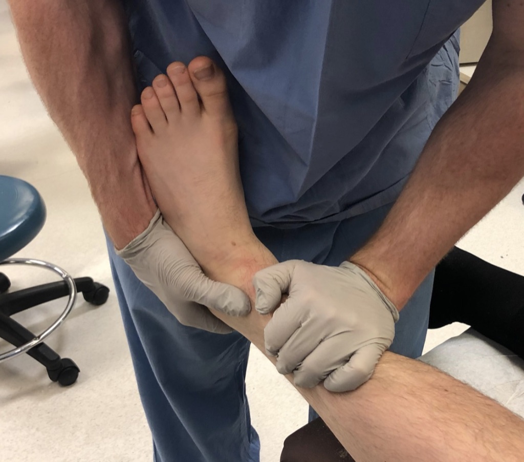
Pull the calcaneus anteriorly while pushing posteriorly on the tibia
-It is often beneficial to place the foot into significant plantar flexion in order to assist reduction
Reduction is often verified by a satisfying clunk and restoration of more normal contour of the ankle
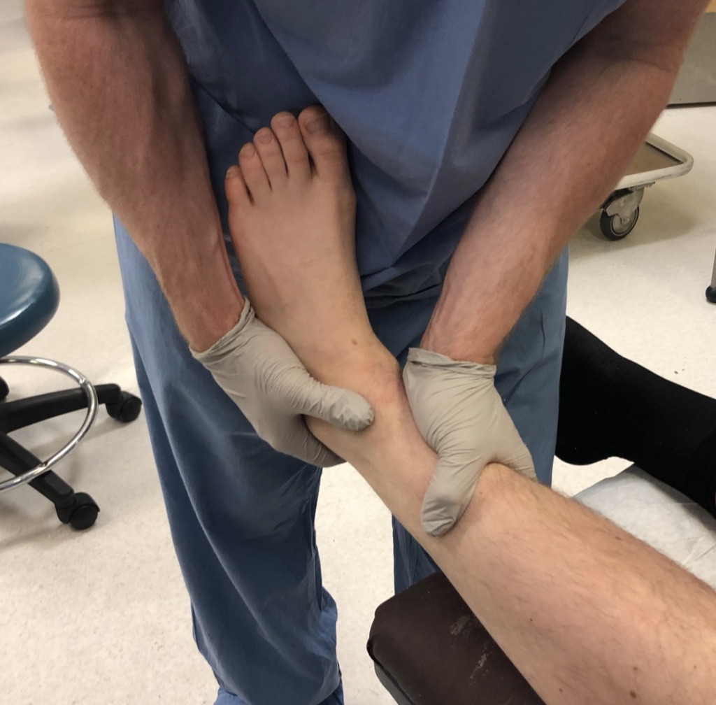
Once talus is reduced beneath the tibia, proceed to reducing the displacement of the fractures
Place one hand on the anteromedial leg above the medial malleolus and one hand on the posterolateral heel
Direct the medial hand laterally and slightly posteriorly
Direct the posterolateral hand anteromedially
Dip and apply splint Mold splint to counteract the deforming force (same positions and forces as just described)
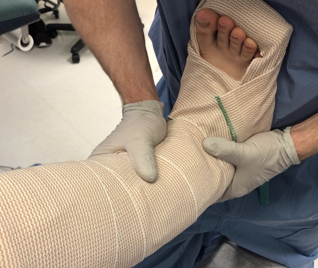
If the Ankle is not too unstable, aim to place the ankle in neutral position to prevent equinus contracture. Dorsiflexion can cause the talus to dislocate posteriorly, so be thoughtful and conscientious about trying to achieve neutral dorsiflexion in an unstable ankle.
Make sure to consistently hold the mold on your splint, repetitive molding and relaxing will both longer for the plaster to set and will fatigue the plaster.
Do not stop holding mold until the plaster is fully set
Post reduction imaging and protocols
Imaging: Post-reduction AP, Mortise, and Lateral of the Ankle in XR suite
CT AFTER acceptable post-reduction ankle radiograph
Restrictions: NWB, Maintain splint
Dispo: if a reasonable person who will be safe at home on a pair of crutches, discharge with close follow-up for skin check and discussion of surgery. Send out on ASA 325mg daily for DVT prophylaxis
If unable to safely discharge home, admit for surgical fixation
Pearls & Pitfalls
Potential complications:
Inadequate reduction requiring repeat reduction
Tips for efficiency: After the reduction is done and the ankle is splinted, go to XR and ask if it is OK to bring patient. Then wheel patient there and watch XRs yourself. This way if the reduction is poor, you can re-reduce under the same hematoma block.